SCAPHO-LUNATE DISSOCIATION
(SL LIGAMENT INJURY)
our website is for educational purposes only. the information provided is not a substitution for seeing a medical doctor. for the treatment of a medical condition, see your doctor. we update the site frequently but medicine also changes frequently.
What is a Scapho-Lunate Dissociation?

A Scapho-Lunate Dissociation is a injury in your wrist. Two of your wrist bones, the Scaphoid and the Lunate, drift apart when an important ligament is torn (the SL Ligament).
Our ligaments connect one bone to another. They keep our bones in alignment and they prevent our bones from wobbling all over the place. Whenever two bones meet up they form a joint, and its the ligaments that hold the joint together and prevent dislocations.
The SL ligament can be torn when someone falls onto an outstretched hand and hyper-extends their wrist.
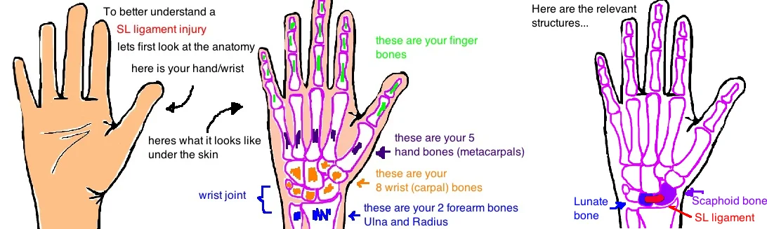
The Scaphoid and Lunate are two of your wrist bones (there are a total of 8 small wrist bones, called carpal bones, that allow for a lot of wrist and hand mobility). The scaphoid and lunate are neighbors and both have seats in the distal radius and together form the radiocarpal joint (wrist joint). They are held together by the Scapho-Lunate Ligament, aka SL ligament (which consists of a weak ligament in the front and a very strong ligament in the back of the wrist).
This ligament is critical for the stability of the whole wrist because it holds the front row of wrist bones in a normal position. If the ligament is torn, the wrist bones (especially the Lunate) will bend backwards (called a DISI deformity) and arthritis will develop over time.
The Scapho-Lunate Dissociation can be an isolated injury or it can be part of a bigger injury that involves multiple ligaments in the wrist being torn. When multiple ligaments are torn, they follow a classic pattern, described by the Mayfield Classification, whereby the energy of a trauma to the wrist travels around the Lunate bone causing "Perilunate" injury. If these injuries are not treated, the wrist bones become unstable (like a bag of marbles) and pain, weakness and arthritis are likely to develop.
Diagnosing a Scapho-Lunate Dissociation:
People with this injury report pain in their wrist and decreased grip strength.
Diagnosis of this injury can be difficult at times. When the ligament is gone, the two bones drift apart, but they may not do so immediately. Right after the injury, x-rays may appear normal and that’s because there are other ligaments (like the STT and the SC ligament) that hold the scaphoid in place, and so even with the SL ligament torn, theres enough overall ligament support to keep everyone in the right position for the short time.
However, if the hand is x-ray’d while you are gripping a pencil, the grip position will “stress” the SL interval and may cause it to widen. This is called dynamic SL Dissocation. Sometimes multiple ligaments are injured and the SL interval is widened without being stressed. This is called static SL Dissocation. Sometimes dynamic Dissocation can become static dissociation when the other ligaments, which hold the scaphoid in place become loose over time due to the increased strain (like a rubber band losing its spring over time).
To call the interval between the scaphoid and lunate widened, it must be 3 mm wider than the interval on the other hand. The widening, almost looks like a gap between someones front two teeth, thats why this injury on x-ray is called “the Terry Thomas sign” after an old comedian with gapped teeth (now we can think of it as the Michael Straham sign).
Over time the scaphoid flexes forward and you see a cortical ring on xray, you can also look at the lateral x-ray and measure the angle formed by the scaphoid and lunate, it should be 45° but will increase to > 70° as the scaphoid flexes forward.
This injury can also be diagnosed with physical exam, people are tender over the SL interval on the dorsal aspect of their wrist, you may feel a cluck of the scaphoid as the wrist is moved from ulnar to radial (known as the Watson test).
Once a doctor suspects SL ligament injury they can order an MRI (which may show tears) or they can perform an arthroscopy (use a tiny camera to look at the ligaments themselves). The problem with MRI is that these ligaments are tiny, you need a really good quality MRI machine and a really smart radiologist reading the MRI to know whats normal and whats injured.
Treating a Scapho-Lunate Dissociation:
These injuries are treated to prevent wrist arthritis. Without this ligament, the bones wont stay in their normal position and over time this causes arthritis. There are different approaches to treatment depending on the severity of injury. Unfortunately, all of the treatment options are surgical.
Arthroscopy is surgery using a small camera and tiny instruments. Doctors can assess the severity of the SL injury by placing a probe between the two bones and seeing how much separation occurs.
If there is only a partial tear, and the dorsal portion of the SL ligament is intact (the strongest part of the ligament), then a doctor will put a few pins to hold the bones together for 6-8 wks while the ligament heals.
If the dorsal and volar portion of the SL ligament is torn, and the injury is recent, and the SL interval is only widen when stressed, you can sometimes repair the ligament (tie it back together).
But if too much time has passed, the ligament will strivel up and a new ligament needs to be made (doctors borrow one of our tendons, like the FCR, or the palmaris longus tendon).
What is the long term outcome?
So why do doctors fix these injuries? Once the scaphoid and lunate are separated, they like to go in different directions. They have other ligament attachements which pull them elsewhere. The scaphoid flexes forward because of the radio-scapho-capitate ligaments, while the lunate and triquetrium extend backward. This disruption in the proximal row is called a DISI deformity (dorsal intercalated segment instability). This deformity will eventually lead to osteoarthritis of the wrist. Its called a SLAC wrist (Scapholunate advanced collapse) but its really just referring to arthritis that starts with the scaphoid and radial styloid impingement then progress to radio-scaphoid arthritis, then midcarpal arthritis (some of the wrist) and finally pan-carpal arthritis (the whole wrist). These can be painful and need bigger corrective surgeries.
The patients do well after this surgery as long as the correct surgery is chosen.

Reference
1. Berger RA. The gross and histologic anatomy of the scapholunate interosseous ligament. J Hand Surg 1996; 21: 170-78. see article. anatomy overview.
2. Walsh JJ, Berger RA, Cooney WP. Current status of scapholunate interosseous ligament injuries. JAAOS 2002; 10: 32-42. see article. review.
3. Short WH et al. A dynamic biomechanical study of scapholunate ligament sectioning. J Hand Surg 1995; 20: 986-999. see article. without SL lig, scaphoid flex, lunate ext.
4. Mayfield JK et al. Carpal dislocations: Pathomechanics and progressive perilunar instability. J Hand Surg 1980;5:226–241. see article. watson shuck test demonstrates SL instability. also tender just distal to listers tubercle.
5. Watson HK, Weinzweig J, Zeppieri J: The natural progression of scaphoid instability. Hand Clin 1997;13:39–49. see article. describes the SLAC wrist.
6. Weiss AP et al. Comparison of the findings of triple-injection cinearthrography of the wrist with those of arthroscopy. JBJS 1996;78:348–356. see article. wrist arthrography only 60% sensitive compared with arthroscopy.
7. Totterman SM, Miller RJ: Scapholunate ligament: Normal MR appearance on three-dimensional gradient recalled-echo images. Radiology1996;200:237–241. see article. MRI is only questionably helpful with diagnosis, only 40% sensitive compared w. arthroscopy.
8. Geissler WB et a. Intracarpal soft-tissue lesions associated with an intra-articular fracture of the distal end of the radius. JBJS 1996;78:357–365. see article. arthroscopy is best for diagnosing and staging SL dissociation. now considered gold standard.
9. Ruch DS et a. Arthroscopic management of partial scapholunate and lunotriquetral injuries of the wrist. J Hand Surg 1996;21:412–417. see article. Pin partial tears for 6-8 wks to prevent progression. < 3 mo since injury (acute), <3 mm diastasis (dynamic instability).
10. Lavernia CJ, Cohen MS, Taleisnik J: Treatment of scapholunate dissociation by ligamentous repair and capsulodesis. J Hand Surg 1992;17:354–359. see article. acute full tear req. open (dorsal approach) repair. lig is usually attached to lunate.
11. Brunelli GA, Brunelli GR. A new technique to correct carpal instability with scaphoid rotary subluxation: A preliminary report. J Hand Surg 1995;20. see article. tenodesis with FCR.